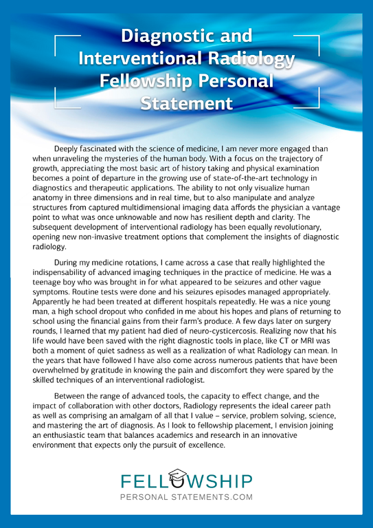
If you want a career as an internist, here is how competitive the medical specialty is to match into an internal medicine residency. Internal medicine is the branch of medicine that involves physicians who provide long-term, comprehensive care in the office and the hospital, managing both common and complex illnesses in adolescents, adults, and the elderly Personal beliefs and medical practice. This page contains the curricula documents for specialty training in clinical radiology. Document downloads. Current curriculum. Clinical radiology curriculum Previous editions. Clinical radiology curriculum Modern slavery statement; Nov 21, · Nearly 95 percent of U.S. allopathic senior medical students matched for postgraduate year 1 (PGY-1) positions in the Main Residency Match, but that’s not to say the competition in certain specialties wasn’t fierce. Find out about the most
How Competitive is an Internal Medicine Residency? | Prospective Doctor
Medical imaging is the technique and process of imaging the interior of a body for clinical analysis and medical intervention, as well as visual representation of the function of some organs or tissues physiology. Medical imaging seeks to reveal internal structures hidden by the skin and bones, as well as to diagnose and treat disease. Medical imaging also establishes a database of normal anatomy and physiology to make it possible to identify abnormalities, radiology personal statement.
Although imaging of removed organs and tissues can be performed for medical reasons, such procedures are usually considered part of pathology instead of medical imaging. As a discipline and in its widest sense, it is part of biological imaging and incorporates radiologywhich uses the imaging technologies of X-ray radiographymagnetic resonance imagingultrasoundendoscopyelastographytactile imagingthermographyradiology personal statement, medical photographynuclear medicine functional imaging techniques as positron emission tomography PET and single-photon emission computed tomography SPECT, radiology personal statement.
Measurement and recording techniques that are not primarily designed to produce imagessuch as electroencephalography EEGradiology personal statement, magnetoencephalography MEGelectrocardiography ECGand others, represent other technologies that produce data susceptible to representation as a parameter graph vs.
time or maps that contain data about the measurement locations. In a limited comparison, these technologies can be considered forms of medical imaging in another discipline. As of5 billion medical imaging studies had been conducted worldwide. Medical imaging is often perceived to designate the set of techniques that noninvasively produce images of the internal aspect of the body.
In this restricted sense, medical imaging can be seen as the solution of mathematical inverse problems. This means that cause the properties of living tissue is inferred from effect the observed signal. In the case of medical ultrasoundthe probe consists of ultrasonic pressure waves and echoes that go inside the tissue to show the internal structure.
In the case of projectional radiographythe probe uses X-ray radiationwhich is absorbed at different radiology personal statement by different tissue types such as bone, muscle, and fat. The term " noninvasive " is used to denote a procedure radiology personal statement no instrument is introduced into a patient's body, which is the case for most imaging techniques used, radiology personal statement. In the clinical context, "invisible light" medical imaging is generally equated to radiology or "clinical imaging" and the medical practitioner responsible for interpreting and sometimes acquiring the images is a radiologist.
Dermatology and wound care are two modalities that use visible light imagery. Diagnostic radiography designates the technical radiology personal statement of medical imaging and in particular the acquisition of medical images.
The radiographer or radiologic technologist is usually responsible for acquiring medical images of diagnostic quality, radiology personal statement, although some radiological interventions are performed by radiologists. As a field of scientific investigation, medical imaging constitutes a sub-discipline of biomedical engineeringmedical physics or medicine depending on the context: Research and development in the area of instrumentation, image acquisition e.
under investigation. Many of the techniques developed for medical imaging also have scientific and industrial applications. Two forms of radiographic images are in use in medical imaging. Projection radiography and fluoroscopy, radiology personal statement, with the latter being useful for catheter guidance. These 2D techniques are still in wide use despite the advance of 3D tomography due to the low cost, high radiology personal statement, and depending radiology personal statement the application, lower radiation dosages with 2D technique.
Radiology personal statement imaging modality utilizes a wide beam of x rays for image acquisition and is the first imaging technique available in modern medicine. A magnetic resonance imaging instrument MRI scanneror "nuclear magnetic resonance NMR imaging" scanner as it was originally known, uses powerful magnets to radiology personal statement and excite hydrogen nuclei i.
Radio frequency antennas "RF coils" send the pulse to the area of the radiology personal statement to be examined. The RF pulse is absorbed by protons, radiology personal statement, causing their direction with respect to the primary magnetic field to change. When the RF pulse is turned off, the protons "relax" back to alignment with the primary magnet and emit radio-waves in the process.
This radio-frequency emission from the hydrogen-atoms on water is what is detected and reconstructed into an image. The resonant frequency of a spinning magnetic dipole of which protons are one example is called the Larmor frequency and is determined by the strength of the main magnetic radiology personal statement and the chemical environment of the nuclei of interest. MRI uses three electromagnetic fields : a very strong typically 1.
Like CTMRI traditionally creates a two-dimensional image of a thin "slice" of the body and is therefore considered a tomographic imaging technique. Modern MRI instruments are capable of producing images in the form of 3D blocks, which may be considered a generalization of the single-slice, tomographic, concept.
Radiology personal statement CT, MRI does not involve radiology personal statement use of ionizing radiation and is therefore not associated with the same health hazards, radiology personal statement.
For example, because MRI has only been in use since the early s, there are no known long-term effects of exposure to strong static fields this is the subject of some debate; see 'Safety' in MRI and therefore there is no limit to the number of scans to which an individual can be subjected, in contrast radiology personal statement X-ray radiology personal statement CT.
However, there are well-identified health risks associated with tissue heating from exposure to the RF field and the presence of implanted devices in the body, such as pacemakers. These risks are strictly controlled as part of the design of the instrument and the scanning protocols used.
Because CT and MRI are sensitive to different tissue properties, the appearances of the images obtained with the two techniques differ markedly. In CT, X-rays must be blocked by some form of dense tissue to create an image, so the image radiology personal statement when looking at soft tissues will be poor. Radiology personal statement MRI, radiology personal statement any nucleus with a net nuclear spin can be used, the proton of the hydrogen atom remains the most widely used, especially in the clinical setting, because it is so ubiquitous radiology personal statement returns a large signal.
This nucleus, present in water molecules, allows the excellent soft-tissue contrast achievable with MRI. A number of different pulse sequences can be used for specific MRI diagnostic imaging multiparametric MRI or mpMRI.
It is possible to differentiate tissue characteristics by combining two or more of the following imaging sequences, depending on the information being sought: T1-weighted T1-MRIT2-weighted T2-MRIradiology personal statement, diffusion weighted imaging DWI-MRIdynamic contrast enhancement DCE-MRIand spectroscopy MRI-S. Radiology personal statement example, imaging of prostate tumors is better accomplished using T2-MRI and DWI-MRI than T2-weighted imaging alone.
Nuclear medicine encompasses both diagnostic radiology personal statement and treatment of disease, and may also be referred to as molecular medicine or molecular imaging and therapeutics. Different from the typical concept of anatomic radiology, nuclear medicine enables assessment of physiology.
This function-based approach to medical evaluation has useful applications in most subspecialties, notably oncology, neurology, and cardiology.
Gamma cameras and PET scanners are used in e. scintigraphy, SPECT and PET to detect regions of biologic activity that may be associated with a disease.
Relatively short-lived isotopesuch as 99m Tc is administered to the patient. Isotopes are often preferentially absorbed by radiology personal statement active tissue in the body, and can be used to identify tumors or fracture points in bone. Images are acquired after collimated photons are detected by a radiology personal statement that gives off a light signal, which is in turn amplified and converted into count data.
Fiduciary markers are used in a wide range of medical imaging applications. Images of the same subject produced with two different imaging systems may be correlated called image registration by placing a fiduciary marker in the area imaged by both systems.
In this case, a marker which is visible in the images produced by both imaging modalities must be used. By this method, functional information from SPECT or positron emission tomography can be related to anatomical information provided by magnetic resonance imaging MRI. Medical ultrasound uses high frequency broadband sound waves in the megahertz range that are reflected by tissue to varying degrees to produce up to 3D images.
This is commonly associated with imaging the fetus in pregnant women. Uses of ultrasound are much broader, radiology personal statement, however. Other important uses include imaging the abdominal organs, heart, breast, muscles, tendons, arteries and veins, radiology personal statement. While it may provide less anatomical detail than techniques such as CT or MRI, it has several advantages which make it ideal in numerous situations, in particular that it studies the function of moving structures in real-time, emits no ionizing radiationand contains speckle that can be used in elastography.
Ultrasound is also used as a popular research tool for capturing raw data, that can be made available through an ultrasound research interfacefor the purpose of tissue characterization and implementation of new image processing techniques.
The concepts of ultrasound differ from other medical imaging modalities in the fact that it is operated by the transmission and receipt of sound waves.
The high frequency sound waves are sent into the tissue and depending on the composition of the different tissues; the signal will be attenuated and returned at separate intervals. A path radiology personal statement reflected sound waves in a multilayered structure can be defined by an input acoustic impedance ultrasound sound wave and the Reflection and transmission coefficients of the relative structures.
It is also relatively inexpensive and quick to perform. Ultrasound scanners can be taken to critically ill patients in intensive care units, avoiding the danger caused while moving the patient to the radiology department. The real-time moving image obtained can be used to guide drainage and biopsy procedures. Doppler capabilities on modern scanners allow the blood flow in arteries and veins to be assessed.
Elastography is a relatively new imaging modality that maps the elastic properties of soft tissue. This modality emerged in the last two decades. For example, cancerous tumours will often be harder than the surrounding tissue, and diseased livers are stiffer than healthy ones. The wide clinical use of ultrasound elastography is a result of the implementation of technology in clinical ultrasound machines.
Photoacoustic imaging is a recently developed hybrid biomedical imaging modality based on the photoacoustic effect. It combines the advantages of optical absorption contrast with an ultrasonic spatial resolution for deep imaging in optical diffusive or quasi-diffusive regime.
Recent studies have shown that photoacoustic imaging can be used in vivo for tumor angiogenesis monitoring, radiology personal statement, blood oxygenation mapping, functional brain imaging, and skin melanoma detection, etc.
Tomography is the imaging by sections or sectioning. The main such methods in medical imaging are:. When ultrasound is used to image the heart it is referred to as an echocardiogram. Echocardiography allows detailed structures of the heart, including chamber size, heart function, the valves of the heart, as well as the pericardium the sac around the heart to be seen.
Echocardiography uses 2D, 3D, and Doppler imaging to create pictures of the heart and visualize the blood flowing through each of the four heart valves. Echocardiography is widely used in an array of patients ranging from those experiencing symptoms, such as shortness of breath radiology personal statement chest pain, to those undergoing cancer treatments.
Transthoracic ultrasound has been proven to be safe for patients of all ages, from infants to the elderly, without risk of harmful side effects or radiation, radiology personal statement, differentiating it from other imaging modalities.
Echocardiography is one of the most commonly used imaging modalities in the world due to its portability and use in a variety of applications. In emergency situations, echocardiography is quick, easily accessible, and able to be performed at the bedside, making it the modality of choice for many physicians. FNIR Is a relatively new non-invasive radiology personal statement technique. NIRS near infrared spectroscopy is used for the purpose of functional neuroimaging and has been widely accepted as a brain imaging technique.
Using superparamagnetic iron oxide nanoparticlesmagnetic particle imaging MPI is a developing diagnostic imaging technique used for tracking superparamagnetic iron oxide nanoparticles. The primary advantage is the high sensitivity and specificityalong with the lack of signal decrease with tissue depth, radiology personal statement. MPI has been used in medical research to image cardiovascular performance, radiology personal statement, neuroperfusionand cell tracking. Medical imaging may be indicated in pregnancy because of pregnancy complicationsa pre-existing disease or an acquired disease in pregnancy, or routine prenatal care.
Magnetic resonance imaging MRI without MRI contrast agents as well as obstetric ultrasonography are not associated with any risk for the mother or the fetus, and are the imaging techniques radiology personal statement choice for pregnant women. The amount of data obtained in a single MR or CT scan is very extensive. Some of the data that radiologists discard could save patients time and money, while reducing their exposure to radiation and risk of complications from invasive procedures.
Volume rendering techniques have been developed to enable CT, MRI and ultrasound scanning software to produce 3D images for the physician. To produce 3D images, many scans are made and then combined by computers to produce a 3D model, which can then be manipulated by the physician.
In diagnosing disease of the viscera of the abdomen, radiology personal statement, ultrasound is particularly sensitive on imaging of biliary tract, urinary tract and female reproductive organs ovary, fallopian tubes.
Radiology Personal Statement
, time: 0:36
Medical imaging is the technique and process of imaging the interior of a body for clinical analysis and medical intervention, as well as visual representation of the function of some organs or tissues ().Medical imaging seeks to reveal internal structures hidden by the skin and bones, as well as to diagnose and treat blogger.coml imaging also establishes a database of normal anatomy and Personal beliefs and medical practice. This page contains the curricula documents for specialty training in clinical radiology. Document downloads. Current curriculum. Clinical radiology curriculum Previous editions. Clinical radiology curriculum Modern slavery statement; Nov 21, · Nearly 95 percent of U.S. allopathic senior medical students matched for postgraduate year 1 (PGY-1) positions in the Main Residency Match, but that’s not to say the competition in certain specialties wasn’t fierce. Find out about the most

No comments:
Post a Comment